VATS (Video-Assisted Thoracic Surgery) Explained
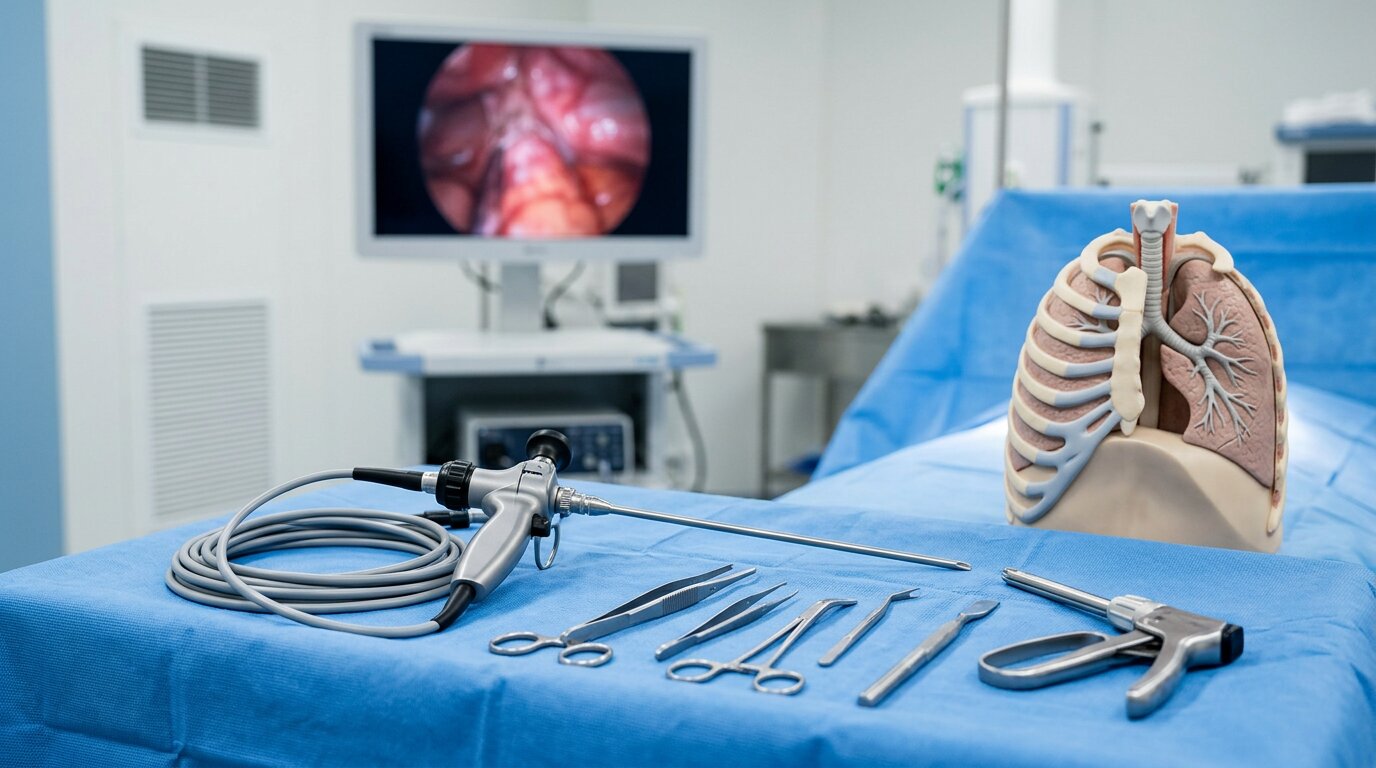
If your doctor’s mentioned video-assisted thoracic surgery (VATS), you’re probably wondering what it really involves and how it might affect your life. You’ll hear that it uses small incisions and a camera instead of opening the chest, but that’s only part of the story. To decide if it’s right for you, you’ll want to know how it compares to traditional surgery, what happens on the day, and what recovery actually looks like.
What Is Video-Assisted Thoracic Surgery (VATS)?
During video-assisted thoracic surgery (VATS), surgeons operate inside the chest using a small camera and specialized instruments instead of creating one large opening between the ribs.
A thin tube called a thoracoscope, along with other instruments, is inserted through several small incisions between the ribs.
The camera provides high-definition images on a monitor, allowing the surgeon to see the chest structures and perform the procedure with precision.
The procedure is done under general anesthesia, and you're positioned on your side opposite the lung being operated on.
Using VATS, the surgeon may remove diseased lung tissue, which can include a small, wedge-shaped section of lung, an entire lobe (lobectomy), or, less commonly, an entire lung (pneumonectomy).
Nearby lymph nodes may also be sampled or removed to assist with diagnosis, staging, or treatment of conditions such as lung cancer or infections.
Because VATS is less invasive than traditional open thoracotomy, many patients experience shorter hospital stays, reduced pain, and faster recovery times. However, surgeons still carefully evaluate each patient’s anatomy and underlying condition before determining whether minimally invasive surgery is appropriate.
In some thoracic cases, surgeons may also evaluate nearby chest wall structures if symptoms involve localized chest pain or inflammation. For example, conditions involving the lower sternum may occasionally require assessment or xiphoid process treatment, particularly when pain around the xiphoid area mimics cardiac or upper abdominal symptoms. Although this is separate from lung surgery itself, chest wall disorders can sometimes overlap with thoracic evaluations, especially when patients present with persistent anterior chest discomfort.
VATS vs Open Chest Surgery: Key Benefits and Risks
While both approaches treat similar chest conditions, video-assisted thoracic surgery (VATS) differs from traditional open chest surgery mainly in how the surgeon accesses the lung and surrounding structures. VATS uses a camera (thoracoscope) and several small incisions between the ribs, which generally reduces the need to widely spread the ribs.
Compared with an open thoracotomy, VATS is usually associated with less postoperative pain, fewer complications, and a shorter hospital stay, often around one to three days. Many patients resume normal activities more quickly and can begin any needed follow‑up treatments sooner. However, VATS still carries risks, including pneumonia, bleeding, injury to nearby nerves or organs, and complications related to anesthesia. The choice between VATS and open surgery depends on factors such as the specific disease, its extent, the patient’s overall health, and the surgeon’s expertise.
When Do Doctors Use VATS and Who Is It For?
Doctors use video‑assisted thoracic surgery (VATS) when chest conditions can be diagnosed or treated safely through small incisions rather than a large open chest incision.
It's commonly used for early‑stage lung cancer (usually stage 1–2) to remove tumors or obtain tissue to confirm the diagnosis.
VATS may also be used after chemotherapy or radiation has reduced the size of a tumor, making a minimally invasive approach more feasible.
In addition, VATS is used to biopsy or manage pleural effusions, mesothelioma, and other diseases affecting the pleura or lungs.
Suitability for VATS depends on several factors, including overall health, lung function, the type and stage of cancer or other condition, and the availability of a thoracic surgeon with experience in minimally invasive techniques.
How to Prepare for VATS and What to Expect on Surgery Day
Before your VATS procedure, your care team will review a clear plan so you understand how to prepare and what to expect on the day of surgery. You'll usually need to avoid eating and drinking for several hours beforehand and follow specific instructions about which prescription medications, over‑the‑counter drugs, vitamins, and herbal supplements to stop or adjust.
Before surgery, you may have blood tests, lung function tests, and heart evaluations such as an EKG. Imaging studies, including a chest X‑ray, CT scan, or PET scan, may be done to help guide the surgical approach.
On the day of surgery, you'll receive general anesthesia and a breathing tube will be placed to support your breathing. You'll typically lie on the side opposite the lung being operated on.
The surgeon will make several small incisions between your ribs to insert the camera and instruments. The procedure usually lasts about 2–3 hours.
At the end of surgery, a chest tube is commonly left in place temporarily to help drain air and fluid from the chest and allow the lung to re‑expand.
Recovery After VATS: Timeline, Activity Limits, and When to Call Your Doctor
After VATS, recovery continues in stages.
Most people stay in the hospital for about 1–2 nights, during which time they begin deep-breathing exercises, coughing, and walking with assistance.
The chest tube is usually removed before discharge, once the lung is expanding well and drainage has decreased.
At home, recovery commonly takes about 2–8 weeks, depending on the specific procedure, your overall health, and any complications.
Activity is increased gradually: walking and light daily tasks are encouraged, while heavy lifting, vigorous exercise, and activities that cause significant pain are typically restricted until your surgeon provides clearance.
Pain is managed with prescribed medications, and some patients may be referred for breathing exercises or physical therapy to support lung function and mobility.
You should contact your healthcare team promptly if you develop worsening shortness of breath, fever or chills, increasing or new chest pain, redness or drainage at the incision sites, or any other sudden change that concerns you, as these may indicate infection, lung problems, or other complications.
Conclusion
VATS gives you a way to treat many chest and lung problems with smaller incisions, less pain, and a faster return to your routine. You’ll still need to prepare carefully, follow your care team’s instructions, and give your body time to heal. Ask questions, understand your risks and options, and speak up about any symptoms after surgery. By staying informed and involved, you can play an active role in your recovery and long‑term lung health.
|








